
Organoids
Organoids are miniaturized, three-dimensional structures grown from stem cells that mimic the complexity and function of real organs. They are used in research to study disease, drug responses and organ development in a controlled laboratory environment.
Organoids
Organoids are miniaturized, three-dimensional structures grown from stem cells that mimic the complexity and function of real organs. They are used in research to study disease, drug responses and organ development in a controlled laboratory environment.
Organoids
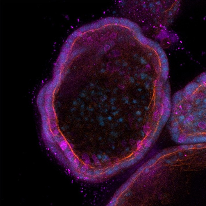
At our Institute, the integration of organoids, 3D printing technologies, and biomimetic models is opening new frontiers in biomedical research. These innovative approaches offer the possibility to create more accurate and physiologically relevant in vitro models, accelerating the development of personalized therapies and reducing the need for animal experimentation. Collaboration across different disciplines, from cell biology to materials engineering, is proving fundamental to addressing the complex challenges of modern medicine, promising significant advances in the understanding and treatment of numerous diseases.
Biomimetic Models and Applications in Hematology
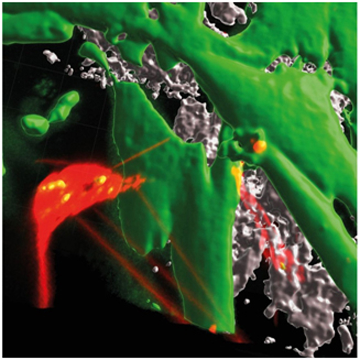
Prof. Martina Pigazzi, PI of the Target discovery and biology of acute myeloid leukemia has developed a 3D biomimetic model of the leukemic bone marrow niche in collaboration with Dr.ssa Monica Sandri lab (ModEls Lab – Biomimetic Composite Materials for Health ISSMC-CNR). This scaffold, composed of hydroxyapatite nanocrystals and collagen (70/30 wt%), faithfully reproduces the composition of bone trabeculae in the marrow niche and allows for long-term co-cultures of mesenchymal stromal cells and leukemic blasts. The model has been further implemented with the addition of endothelial cells, Schwann cells, and monocytes, offering an advanced platform for studying leukemia pathogenesis and drug response both in vitro and in vivo (Borella G. et al, Blood 2021).
Heterotypic tunneling nanotubes that interconnected acute myeloid leukemia (AML) cells (red, Vybrant-DiI) and mesenchymal stromal cells derived from the bone marrow of an AML pediatric patient at diagnosis (green, Calcein-AM) in 3D in vitro leukemia bone marrow niche model (grey correspond to the collagen and hydroxyapatite of the scaffold).
3D Models for Complex Diseases
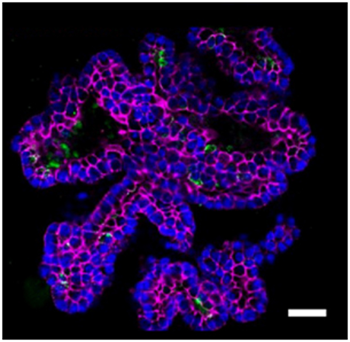
The Stem Cells and Regenerative Medicine Laboratory (PI Prof. Michela Pozzobon) develops various 3D models for complex diseases, including colon organoids for inflammatory bowel disease, lung organoids to simulate inflammatory and oxidative damage, a 3D decellularized human muscle model to study muscle regeneration, and a hydrogel-based model for rhabdomyosarcoma. These models enable the study of cell–matrix interactions and the effect of extracellular vesicles secreted by mesenchymal cells.
Representative image of lung organoid: alveoli with secretory cells (MUC5ac), nuclei (DAPI), and in red phalloidin (cytoskeletal actin).
Neuromuscular Organoids
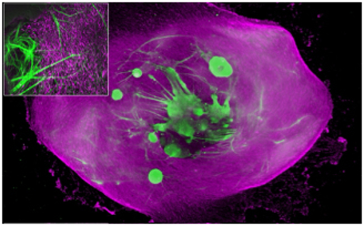
Dr. Anna Urciuolo, PI of the Neuromuscular Engineering Laboratory, develops neuromuscular organoids (NMO) derived from human induced pluripotent stem cells (hiPSC). These models, including 2D cell cultures, self-assembled 3D NMOs and scaffold-engineered NMOs, allow the study of the integrated human neuromuscular system in health, congenital and acquired diseases, offering a platform for high-resolution imaging, omics, and functional analyses.
Whole mount immunofluorescence image showing a tissue-engineered neuromuscular organoid derived from hiPSCs, with integrated muscular (magenta) and neuronal (green) compartments.
Tumor Organoids and 3D Models for Cancer Research
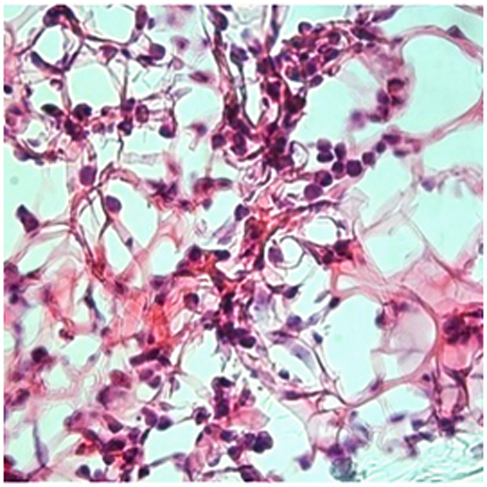
The Transcriptomics and Functional Genomics Laboratory in JMML and B-ALL (PI Dr. Silvia Bresolin) has developed a 3D in vitro system called the “atypical organoid” for juvenile myelomonocytic leukemia (JMML). This model supports long-term proliferation and survival of JMML progenitor cells, overcoming the limitations of conventional 2D cultures and providing a novel tool for studying this rare pediatric disease.
Hematoxylin/eosin staining of fixed pd-JAO 3D structure. Notably, JMML cells are incorporated in the extracellular matrix. Original magnification 40×. pd-JAO: patient-derived JMML Atypical Organoid; JMML: juvenile myelomonocytic leukemia. Adapted from Cani A. et al., Blood Adv (2023) 7 (8): 1513–1524.
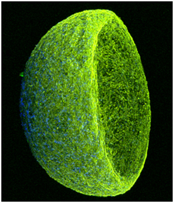
Dr. Luca Persano (PI of the Biology of CNS Pediatric Tumors Laboratory) generates brain tumoroids derived from primary glioblastoma, pediatric medulloblastoma, and meningioma cells. These 3D models replicate tumor heterogeneity and tissue organization of the original tumors, demonstrating their potential for diagnostic, mechanistic, and personalized medicine studies. Interestingly, heterotypic glioblastoma tumoroids – incorporating a normal brain cells component – are under development.
3D reconstruction of a 30 day-cultured brain tumoroid generated from GFP-expressing patient-derived cells.
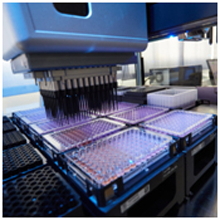
The Organoid Facility hosts advanced high-throughput screening (HTS) platforms based on physiologically relevant 3D cell culture models. Prof. Giampietro Viola, PI of the Experimental Pharmacology Laboratory, together with Dr. Elena Mariotto (Junior PI of the High-Throughput Drug Screening for Precision Oncology Unit), developed and optimized a screening workflow using tumor spheroids derived from pediatric brain cancers. Compared with conventional 2D cultures, 3D spheroid models more accurately reproduce key features of tumor biology, including cellular architecture, metabolic gradients, drug penetration, and treatment response. Using these models, the platform has enabled the identification of metabolic and pharmacological vulnerabilities that are not detectable in standard in vitro systems. The platform integrates automated liquid handling and multi-condition screening in 96-well plate format, allowing the parallel evaluation of hundreds of compounds or experimental conditions on 3D tumor models. Current efforts are also focused on the development of quantitative image-based analysis pipelines to further improve data acquisition and phenotypic profiling. This approach provides a powerful tool for precision oncology studies, drug repurposing programs, and the identification of novel therapeutic strategies for pediatric and adult cancers.
Image: 96-well plate used for high-throughput screening (HTS). This platform enables the simultaneous testing of hundreds of compounds or experimental conditions on 3D cell cultures, such as tumor spheroids. By employing models that more closely resemble the architecture and behavior of real tumors compared to traditional 2D systems, this approach allows for a more accurate identification of candidate compounds for cancer therapy.
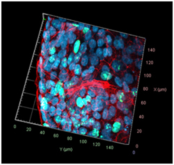
The NanoInspired Biomedicine Laboratory (PI Prof. Marco Agostini) develops tumor organoids (“tumoroids”) derived from patients with gastrointestinal cancers, in particular colorectal cancer. These tumoroids, cultured in hydrogels derived from decellularized extracellular matrix, provide a patient specific preclinical model to evaluate chemotherapy response, opening new perspectives for precision medicine.
Confocal microscopy image of patient-derived colorectal cancer organoids acquired along the X, Y, and Z axes. Ki67 was used as a proliferation marker (green), F-actin for visualization of cytoskeletal microfilaments (red), and DAPI for nuclear counterstaining (blue).